Silver-based black and white gelatine photographs are prone to mirroring (developing a mirror-like sheen), yellowing and fading (see Fig. 1). This is caused by pollutants, high humidity and residual processing chemicals (sodium or ammonium thiosulfate).
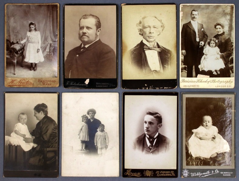
Fig. 1 A selection of silver-based photographs with different degrees of yellowing
Experts claim that they can diagnose the cause of deterioration based on the degree and hue of yellowing. For example, a photograph might appear red-brown due to residual processing chemicals or yellow-orange due to the effects of humidity. This is attributed to changes in the size and shape of the image silver.
I wanted to find out more. Could a subjective colour assessment, which requires many years’ experience, be quantified using a colour measurement? With the help of a Konica Minolta spectrophotometer CM-2600d, I decided to look at the effects of humidity and residual processing chemicals on a set of artificially aged photographs, some of which had been processed well and others poorly.

Fig. 2 A set of laboratory samples before and after ageing. Top row, left to right: well-processed un-aged, aged 50 °C/70% RH, and aged 50 °C/90% RH. Bottom row, left to right: insufficiently washed un-aged, aged 50 °C/70% RH, and aged 50 °C/90% RH.
Fig. 2 shows a set of laboratory samples. I aged them at 50 °C/70% RH and 50 °C/90% RH respectively. The greatest colour contrast was visible after ageing at 90% RH, when the poorly processed photographs looked more purplish, while the well-processed ones looked more yellow. It was more difficult to distinguish between the well-processed sample aged at 50 °C/90% RH and the poorly processed sample aged at 50 °C/70% RH with the naked eye. The colour measurements could do this more effectively.

Fig. 3 The colour differences (ΔL* Δa* Δb* values) for the well-processed and insufficiently washed laboratory samples. Top left: well-processed aged at 50 °C/70% RH. Top right: well-processed aged at 50 °C/90% RH. Bottom left: insufficiently washed aged at 50 °C/70% RH. Bottom right: insufficiently washed aged at 50 °C/90% RH.
The bar charts (see Fig. 3) are colour coded to show the differences in the hue of the photograph before and after ageing using L* (dark to light), a* (green to red), and b* (blue to yellow). On the whole, the well-processed photographs became more yellow while those that had been poorly processed became redder and darker. This proved that in a limited sample set a colour measurement could diagnose the causes of yellowing more effectively than the naked eye.

Fig. 4 Transmission electron microscopy pictures of silver filaments x150k (scale bar measures 100nm). Top row, left to right: well-processed un-aged, aged 50 °C/70 % RH, and aged 50 °C/90 % RH. Bottom row, left to right: insufficiently washed un-aged, aged 50 °C/70 % RH, and aged 50 °C/90 % RH.
I then looked at the silver using a transmission electron microscope. Fig. 4, taken at x150,000, shows how the corresponding silver particles have changed shape and size. The well-processed photographs generated more colloidal silver (tiny spherical silver particles) and became more fragmented, while the sulphur-affected photographs produced less colloidal silver and became more homogenous. This corresponded to the perceived colours: the well-processed photographs were more yellow whilst the poorly washed photographs were redder.

Fig. 5 Historical samples. Clockwise from top left: ‘Aeroplane’, ‘Ceremony’, ‘Street Scene’ and ‘Crumpled Manuscript’. Note: these photographs have been used for a separate ageing experiment and the discoloured rectangles can be disregarded.
I then looked at the complexities of applying these findings to historical photographs (see Fig. 5). The behaviour of the silver particles is similar to the laboratory samples, but they come in a much greater variety of sizes and shapes (see Fig. 6). The images on the bottom row of Fig. 6 show the cross section of each photograph. The clustered particles on the right are the baryta layer, while the silver particles are dispersed to their left. The shaded areas are creases in the cross sections.

Fig. 6 Transmission electron microscope images of the historical samples. Left to right: ‘Street Scene’, ‘Ceremony’, ‘Aeroplane’ and ‘Crumpled Manuscript’. Top row x150k, bottom row x10k (scale bar measures 100nm).
The range of paper, image tones and processing methods of the historical samples makes direct comparisons with the naked eye challenging. Does the photograph have white highlights or is it yellowed overall? Has it been processed to have a reddish hue or has it been poorly washed? I decided to take colour measurements from a range of image tones in each photograph, including the highlights. The reflectance spectra were able to corroborate the results of a residual sulphur test. ‘Ceremony’, a poorly washed historical photograph, had greater red reflectance than ‘Street Scene’, a well-processed yellowed photograph (see Fig. 7).

Fig. 7 The reflectance spectra of the dark and medium tones of ‘Street Scene’ and ‘Ceremony’. The spectra show an increased red reflectance in the sulphur-affected ‘Ceremony’.
My plan is to experiment on a much larger sample set. If the pattern holds true, then conservators and collection managers would be able to establish causes of deterioration using a non-destructive colour measurement, which could also be adapted for use by volunteers and other non-specialists involved in surveying.
Jacqueline Moon completed a Master of Research in Heritage Science at University College London in 2015, in order to understand the yellowing of silver gelatine photographs. She is now the Senior Conservation Manager for Public and Academic Engagement at The National Archives, and Chair of the Institute of Conservation Photographic Materials Group. You can read more about her work here: https://doi.org/10.1186/s40494-017-0159-9.
